Clinical Scenario
It’s a busy day in ED this morning.
The first patient refers dyspnoea, he has an advanced pulmonary neoplasia, ultrasound and chest x-ray confirm that the left zone is occupated by a pleural effusion. The thoracentesis removes about 1.5 liter of fluid, the patient breathes easily.
The second patient is 65 yo, he has pneumonia, he is septic, it’s impossible to find a vein, the right internal jugular vein is identified by ultrasound and the catheterization is performed without any problem.
Conclusion
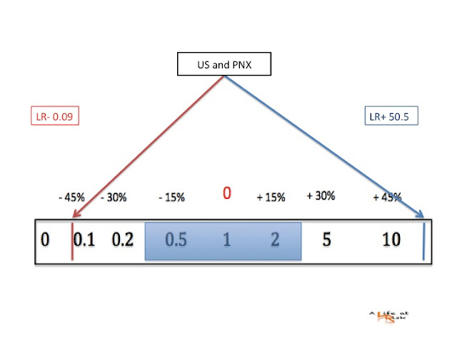
It is a common practice to perform a chest x-ray after procedural thoracic manoeuvres like pleural aspiration or central line inserction. The commonest complication of these tecniques is a PNX. The BTS pleural disease guidelines 2010 say that the probability of this complication after a pleural aspiration using ultrasound guidance is about 2-3%, the same is described for an US-guided vascular access.
Ultrasound compared with a semierect or supine chest x-rays is more accurate for ruling out a PNX.
Enjoy ultrasound!!!!
Bibliography
T. Havelock et al.
Pleural procedures and thoracic ultrasound: British Thoracic Society pleural disease guideline 2010
Thorax 2010; 65 (Suppl 2) ii61-ii76.
A.S.Raja et al.
How accurate is ultrasonography fpr excluding pneumothorax?
Ann Emerg Med vol 61 n 2 Febr 2013
Ciro Paolillo


No comments:
Post a Comment